The human body has long fascinated scientists, artists, and the curious alike. A striking set of images recently caught my attention, blending detailed anatomical models with a glimpse of a historical figure whose life defied medical norms. These visuals—featuring sagittal head sections, elongated spines, and a familiar profile—hint at a rich tapestry of medical history and human anomaly. Let’s explore what these images might represent, with a focus on their potential connection to Joseph Merrick, the “Elephant Man,” and the anatomical studies that followed.
The Anatomical Models: A Window into the Past
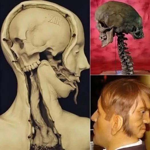
The images include two remarkable anatomical models. The first, a sagittal section of a human head and neck, reveals the brain, skull, and upper spine with the marking “XII” etched into the bone. This style echoes the wax anatomical models crafted in the 18th and 19th centuries, notably by Italian artist Clemente Susini. These models, used in medical schools across Europe, offered a hands-on way to study human anatomy when dissection was limited. The “XII” could denote a specimen number or a reference to the 12th cranial nerve, the hypoglossal, which controls tongue movement—a detail that might tie into studies of speech or deformity.
The second model showcases a preserved skull with an exaggeratedly elongated spine, suggesting either a natural condition or an artistic exaggeration. Such displays were common in medical museums or sideshows, where the unusual was both studied and sensationalized. These models likely served as tools to educate or intrigue, reflecting the era’s blend of science and spectacle.
The Elephant Man: Joseph Merrick’s Story
The profile in the bottom right image bears a striking resemblance to Joseph Merrick, born in 1862 in Leicester, England. Afflicted with severe deformities—attributed to Proteus syndrome or possibly neurofibromatosis—Merrick became known as the “Elephant Man” after being exhibited in Victorian freak shows. His condition caused overgrown bone and soft tissue, particularly on his face and limbs, making him a subject of both pity and fascination.
Rescued from exploitation by surgeon Sir Frederick Treves in 1886, Merrick spent his final years at the London Hospital. His case offered a rare opportunity to study such a condition up close, though he was never fully diagnosed due to the limitations of 19th-century medicine. After his death in 1890 at age 27, his skeleton was preserved for study, and his story inspired countless medical and cultural explorations. The anatomical models in the images could be artistic interpretations or educational tools inspired by Merrick’s unique physiology.
Bridging Science and Spectacle
The combination of these images suggests a bridge between scientific inquiry and public curiosity. In the 19th century, anatomy was both a rigorous discipline and a public draw. Museums like the Hunterian in London displayed preserved specimens, while waxworks and illustrations brought the inner body to life. Merrick’s case, with its visible deformities, fit this dual role—studied by doctors yet gawked at by crowds. The elongated spine model might exaggerate his condition for effect, a common practice to highlight medical rarities.
These visuals also reflect the era’s attempt to understand the human form through art and science. The detailed craftsmanship of the models indicates a dedication to accuracy, while their preservation hints at their value as historical artifacts. Today, they serve as a reminder of how far medical knowledge has come, even as they evoke the humanity behind the science.
The Legacy Today
Joseph Merrick’s story endures, not just as a medical curiosity but as a symbol of resilience. His preserved skeleton remains at the Royal London Hospital, studied ethically to advance understanding of rare conditions. The anatomical models, whether directly linked to him or not, represent a time when such studies pushed the boundaries of knowledge, often with a mix of reverence and exploitation.
Modern medicine has benefited from these early efforts, using advanced imaging and genetics to diagnose conditions like Proteus syndrome with greater precision. Yet, the images retain their power to intrigue, blending the eerie beauty of anatomical art with the human story behind it.
Conclusion
These images—featuring anatomical models and a nod to the Elephant Man—offer a fascinating glimpse into the intersection of science, history, and human curiosity. From the detailed waxworks of the past to Merrick’s poignant legacy, they remind us of our ongoing quest to understand the body’s mysteries. Whether you see them as educational tools or relics of a bygone era, they invite us to reflect on how far we’ve come—and how much more there is to learn.